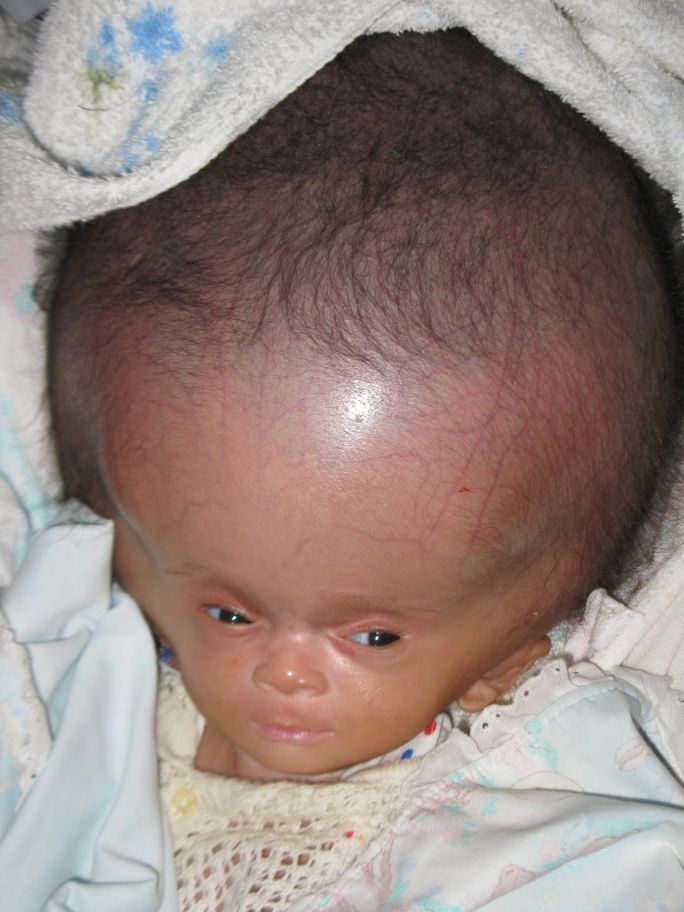

This condition often requires surgical management and helmet therapy may be used. When plagiocephaly occurs with craniosynostosis, the skull deforms due to premature fusion of the sutures in the skull.positional plagiocephaly without craniosynostosis. plagiocephaly followed by craniosynostosis (a birth defect in which the bones in a baby's skull fuse prematurely) and 2. twins, triplets etc), prematurity, congenital muscular torticollis, position of the head and lying in the same position for prolonged periods. It has a number of potential causes, including: first birth, assisted labour, multiple births (e.g. Positional plagiocephaly is an asymmetric deformation of the skull. J Neuroimaging 2017 27(2):162–209.Positional plagiocephaly is increasingly common in infants. Neuroimaging Findings in Pediatric Genetic Skeletal Disorders: a Review. Wagner MW, Poretti A, Benson JE, Huisman TA.Craniosynostosis: Updates in Radiologic Diagnosis. D’Apolito G, Colosimo C, Cama A, Rossi A.Misshapen heads in babies: position or pathology? Ochsner J 2001 3(4):191–199. Craniosynostosis: imaging review and primer on computed tomography. Badve CA, Mallikarjunappa MK, Iyer RS, Ishak GE, Khanna PC.In the online presentation, the anatomy and embryology of the skull and the main craniosynostoses are presented, as well as the protocol for low-dose CT. Postoperative complications such as stroke, hemorrhage, cerebrospinal fluid leaks, intracranial collections, and venous damage or thrombosis must be ruled out. In addition, craniovertebral junction anomalies and signs of increased intracranial pressure need to be assessed carefully. The radiology report should include a section about the vascular anatomy, including the course of the venous sinuses, large scalp or bridging veins (relevant for surgical planning), venous thrombosis, and arterial variants. The neuroradiologist’s role in pediatric assessment is the evaluation of the head shape with regard to premature fusion of the sutures, which can be depicted at CT. Type 3 is similar but without the cloverleaf skull. Type 2 manifests with cloverleaf skull, extreme proptosis, and ankylosis or synostosis of elbows. Frequent manifestations include midface hypoplasia, hypertelorism, broad thumbs and toes, brachydactyly, and syndactyly. There are three types, with different degrees of fusion of the coronal and sagittal sutures. Pfeiffer syndrome is caused by mutations in the FGFR1 or the FGFR2 gene. Systemic features include hearing loss due to auditory meatus atresia, orbital defects, and cervical spine fusion.

Craniofacial features are coronal synostosis (the sagittal and lambdoid sutures can also be affected), midface hypoplasia, and exophthalmos. Further musculoskeletal anomalies include syndactyly, vertebral fusion (often at C5-C6), and joint deformities.Ĭrouzon syndrome is caused by mutations of the FGFR2 or FGFR3 gene. The craniofacial findings include coronal synostosis (rarely, cloverleaf skull), midface hypoplasia, and hypertelorism. Apert syndrome is caused by a mutation in the FGFR2 gene. Syndromic synostoses are caused by specific mutations and manifest with multiple systemic alterations. The keys to differentiate lambdoid synostosis from positional plagiocephaly are the trapezoidal shape of the skull and posterior displacement of the ear. It is characterized by occipitoparietal flattening, mastoid bulging, and contralateral occipitoparietal and frontal bossing. The least common type is lambdoid synostosis (posterior plagiocephaly). Orbital features include quizzical orbits and lateral orbital hypoplasia. Triangular or pearlike shape, parieto-occipital bossing, and narrow anterior cranial fossa are characteristic cranial features. Metopic synostosis (trigonocephaly) is less common. Bicoronal synostosis (brachycephaly) manifests with a widened transverse diameter of the skull, harlequin deformity, and hypertelorism. This deformity is called anterior plagiocephaly with ipsilateral exophthalmos, frontal bone flattening, and contralateral bossing. The second most common type is coronal synostosis, which can be unilateral or bilateral, with the former being more common.


 0 kommentar(er)
